
Keratoconus Treatment Singapore
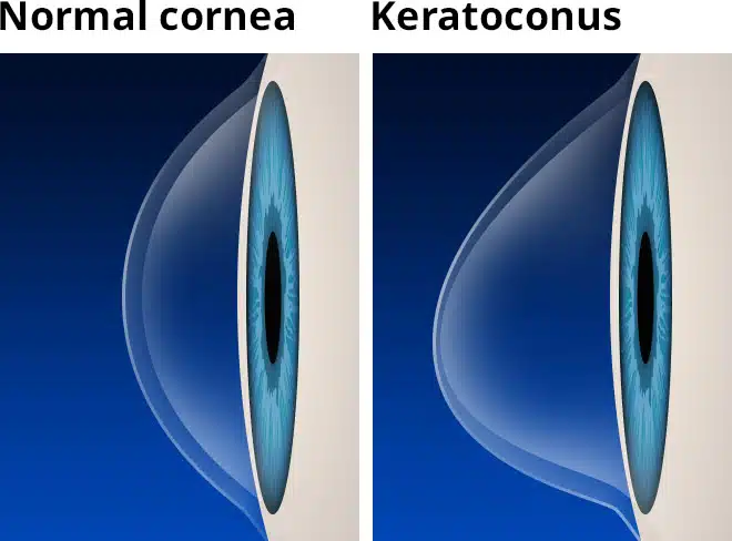
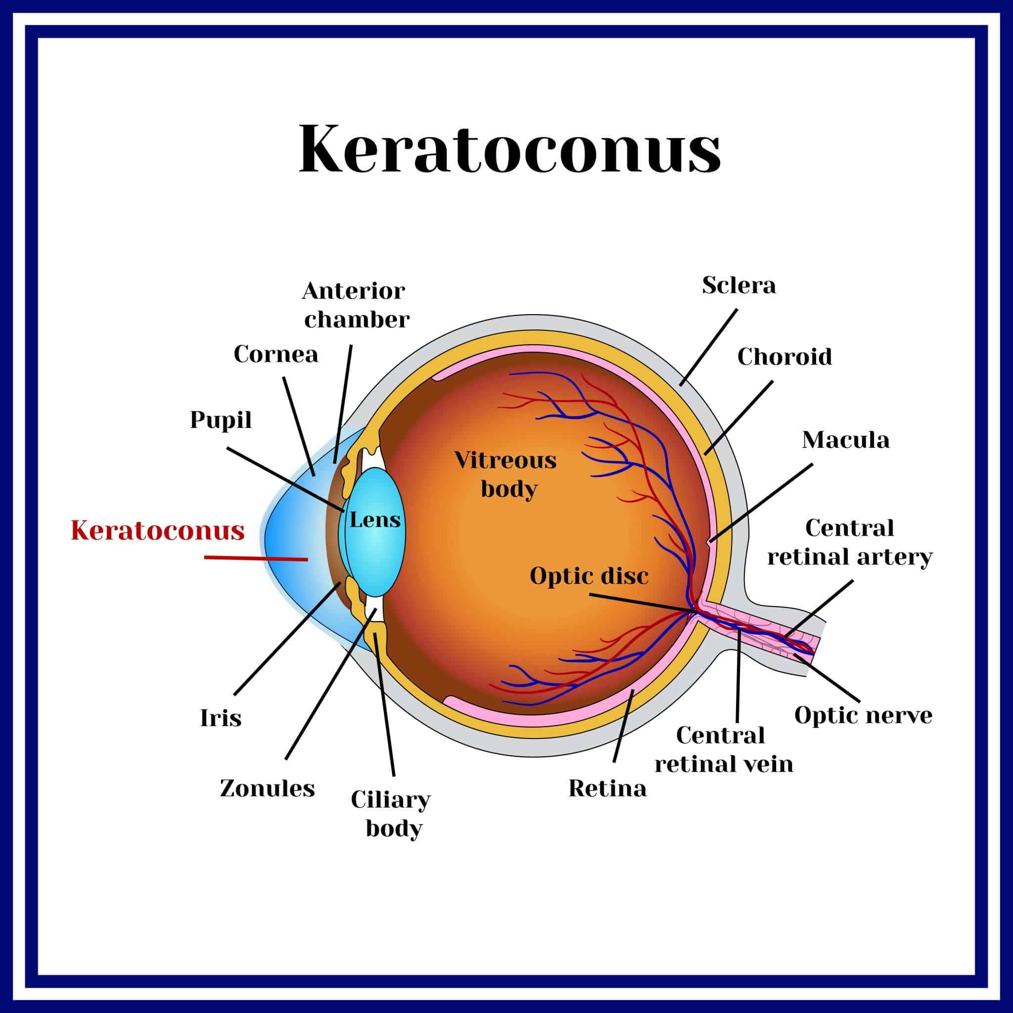
Keratoconus is a progressive eye condition in which the cornea (the clear, dome-shaped front surface of the eye) gradually thins and bulges outward into a cone shape. This change in corneal structure causes light entering the eye to be deflected unevenly, resulting in distorted and blurred vision.
At LSC Eye Clinic, keratoconus is assessed and treated by specialist ophthalmologists with experience in corneal conditions and refractive eye care.
What Is Keratoconus?
In a healthy eye, the cornea maintains a regular, rounded shape that focuses light accurately onto the retina. In keratoconus, the corneal tissue weakens over time, causing it to thin and protrude forward.
This irregular shape disrupts how light is focused, producing blurred, distorted, or doubled vision.
The condition typically presents during the teenage years or early adulthood and can affect one or both eyes. Where both eyes are involved, the degree of change often differs between them.
As the cornea continues to change shape, astigmatism (irregular curvature of the eye’s focusing surface) tends to increase, and glasses or standard soft contact lenses may no longer provide adequate correction.
Causes of Keratoconus
Studies have suggested that an imbalance of enzymes in the cornea exposes the eye to oxidative damage from free radicals. In turn, this damage can lead to the weakening and bulging of the corneal tissue, resulting in keratoconus.
Other factors that could predispose one to keratoconus are:
- Overexposure to the sun’s ultraviolet (UV) rays
- Excessive eye rubbing
- Chronic eye irritation
- Genetics
Why See an Eye Specialist for Keratoconus
Keratoconus does not remain stable in all cases. In some patients, the cornea continues to change over months or years, progressively affecting vision quality and making correction more difficult.
An eye specialist is able to monitor the rate of change through serial corneal mapping, identify when treatment is needed to slow progression, and guide decisions about vision correction options suited to the current severity of the condition.
Routine glasses prescription updates alone do not address the underlying corneal changes. Instead, specialist assessment allows for corneal collagen cross-linking treatment of keratoconus to be considered at an appropriate stage, before significant structural changes occur.
What Are the Signs and Symptoms of Keratoconus?
Symptoms vary depending on the severity and stage of the condition. Early keratoconus may be subtle, and some patients are unaware of the changes until identified during a routine eye examination.
Common signs and symptoms include:
- Distorted or blurred vision: Objects may appear wavy, smeared, or duplicated, particularly at certain distances.
- Frequent prescription changes: A need to update glasses or contact lens prescriptions repeatedly over a short period.
- Ghosting or multiple images: Seeing a shadow or secondary image alongside the main object.
- Increased sensitivity to light and glare: Particularly noticeable at night or in bright environments.
- Difficulty with night vision: Halos or streaking around lights, especially when driving after dark.
- Contact lens discomfort: Lenses may fit poorly or feel unstable as the corneal shape changes.
How Keratoconus Is Diagnosed
Diagnosis involves a combination of vision testing and corneal imaging. Several measurements are taken to assess the shape, thickness, and curvature of the cornea and determine the severity of the condition.
The diagnostic process typically includes:
| Assessment | What It Evaluates |
| Vision testing | Current prescription, including the degree and type of astigmatism |
| Corneal topography and tomography | Surface curvature and overall shape of the cornea, mapped in detail |
| Corneal thickness measurement (pachymetry) | Thickness at multiple points across the cornea |
| Slit-lamp examination | Structural integrity of the cornea and ocular surface health |
Together, these assessments help determine the stage of keratoconus and whether the condition is progressing. They also guide decisions about which treatment options are appropriate for your eyes.
What Is the Treatment for Keratoconus?
Keratoconus vision treatment depends on the severity of the condition and whether it is actively progressing. The goal of treatment is twofold: to correct vision as effectively as possible and, where relevant, to slow the progression of corneal changes.
Glasses
In the early stages, glasses may provide adequate vision correction. As the cornea becomes more irregular, glasses are typically no longer sufficient to achieve clear vision, and contact lens options are usually explored.
Soft Contact Lenses
Soft contact lenses may be suitable in mild keratoconus, where the corneal irregularity is not yet severe. They provide limited correction of irregular astigmatism compared with rigid lens options.
Rigid Gas Permeable (RGP) Lenses
RGP lenses sit on the surface of the cornea and create a smooth optical surface over the irregularity. They provide clearer vision than soft lenses for many patients with moderate keratoconus, though they can take time to adapt to.
Hybrid Lenses
Hybrid lenses combine a rigid central zone with a soft peripheral skirt, providing the optical clarity of an RGP lens with greater comfort. They can be suitable for patients who have difficulty tolerating standard RGP lenses.
Scleral Lenses
Scleral lenses are large-diameter rigid lenses that vault over the entire cornea and rest on the sclera (the white of the eye). The space between the lens and the cornea is filled with saline, creating a smooth refracting surface regardless of the cornea’s shape. They are commonly used in moderate to advanced keratoconus and are often more comfortable than smaller RGP lenses.
Corneal Collagen Cross-Linking (CXL)
Corneal collagen cross-linking (CXL) is a treatment that uses riboflavin eye drops, a type of vitamin B, activated by ultraviolet-A light to strengthen the cornea. Its aim is to stabilise the corneal tissue and slow the progression of keratoconus.
Corneal Transplant
In advanced keratoconus where vision cannot be adequately corrected with lenses, or where the cornea has become significantly scarred, a corneal transplant may be considered. This replaces part or all of the cornea with donor tissue.
Suitability and outcomes vary for every individual. As such, a clinical assessment is required to determine the appropriate treatment approach for your eyes.

When Surgery May Be Needed
For most patients with keratoconus, vision correction with contact lenses and cross-linking to stabilise the cornea are sufficient. However, surgery is considered when these approaches are no longer adequate.
Situations where surgery may be appropriate include:
- Poor vision with contact lenses: When lens-corrected vision no longer meets functional needs.
- Contact lens intolerance: When the irregular corneal surface makes lens wear uncomfortable or impractical.
- Significant corneal scarring: When opacity or scarring reduces vision quality beyond what lenses can address.
- Advanced thinning: When the cornea has become too thin or irregular for other interventions to be effective.
In these situations, a corneal transplant may be considered for treatment. The appropriate surgical approach depends on the depth and extent of corneal involvement.
Recovery and Follow-Up Post Cornea Keratoconus Treatment
Keratoconus requires long-term monitoring, as the condition can continue to evolve over time, even after treatment. A single appointment or intervention is rarely sufficient.
For patients who have had cross-linking, follow-up typically includes corneal mapping at intervals over the first one to two years to confirm that the cornea has stabilised. Contact lens fitting may need to be adjusted as the corneal shape settles.
For patients managed with contact lenses alone, regular reviews allow the clinical team to track changes in corneal curvature and vision and update the lens prescription or type as needed. As dry eye is a common concurrent condition, it may also be monitored during follow-up, and dry eye treatment may be considered as an option.
Where surgery has been performed, recovery depends on the procedure. Patients are advised on specific timelines and restrictions at the time of their consultation.
What to Expect at Your Appointment
A keratoconus assessment at LSC Eye Clinic follows a structured process. Knowing the stages in advance can make the visit feel more straightforward.
Your first visit typically includes the following stages, from initial history-taking through to discussion of treatment options:
- History and Registration: A record of your symptoms, current vision correction, and relevant medical or family history.
- Eye Tests: Vision testing, corneal mapping, corneal thickness measurement, and a slit-lamp examination of the cornea.
- Review of Findings: A discussion of your results and the treatment options suited to your case.
- Next Steps: A possible follow-up appointment to review results or plan treatment.
Patients are encouraged to ask questions about their diagnosis, the likely progression of the condition, and the management options available.
Meet Our Doctor

Dr Daphne Han
Medical Director and Senior Consultant Ophthalmologist
MBBS (Melbourne), MMed (NUS), FRCS (Edinburgh), FAMS (Ophthalmology)
Specialty: Laser Vision Correction and Lens Implant Surgery
Dr Daphne Han is the Medical Director and Senior Consultant Ophthalmologist at LSC Eye Clinic, with over 20 years of clinical experience in ophthalmology. Her subspecialty focus covers laser vision correction and lens implant surgery, with experience in managing corneal and refractive conditions including keratoconus, encompassing LASIK, ZEISS SMILE® pro 2.0, ZEISS PRESBYOND®, Epi-LASIK, LASEK, PRK, and Implantable Collamer Lenses (ICL) for patients unsuitable for laser-based correction.
Dr Han trained at the Singapore National Eye Centre (SNEC), where she served for over 14 years, and has completed advanced fellowship training internationally, including at IROC Zurich under Professor Theo Seiler and at the Cincinnati Eye Institute. She holds an appointment as Adjunct Assistant Professor at Duke-NUS Medical School and remains active as a published author and peer reviewer in the field of refractive surgery.
In addition to refractive procedures, Dr Han performs conventional and laser-assisted cataract surgery and manages conditions including glaucoma and general ophthalmology, providing clinical oversight across a broad range of eye conditions.
Payment Options
We accept cash, NETS, and major credit cards.

Interest-Free Instalment Plans
Available with UOB, OCBC, DBS, and POSB bank credit cards.

MediSave
Keratoconus treatment and other eye procedures are MediSave claimable.
Our clinic is able to assist with your claim.
Many private insurance providers also cover keratoconus treatment surgery claims.

Insurance
We are on the specialist panels of various Insurance Plans:

Why Choose LSC Eye Clinic for Keratoconus Treatment
SPECIALIST-LED CORNEA CARE
Keratoconus assessments and treatments are led by specialist ophthalmologists, giving patients access to thorough clinical evaluation.
90,000+ EYES TREATED SINCE 2005
As an eye clinic in Singapore, LSC Eye Clinic has cared for more than 90,000 eyes, providing patients with clinical depth across a broad range of conditions.
STRUCTURED FOLLOW-UP AND MONITORING
Long-term monitoring is built into the keratoconus care we provide, helping patients track corneal stability and update treatment as needed.
CENTRAL ORCHARD ROAD LOCATION
The clinic at Paragon Medical on Orchard Road offers central access for patients attending assessment and follow-up visits.
Suitability and outcomes vary for every individual. A clinical assessment will determine what keratoconus treatment is appropriate for your eyes.
Frequently Asked Questions About Keratoconus in Singapore
Progression is usually confirmed through serial corneal mapping rather than symptoms alone. If your glasses or contact lens prescription has changed frequently, or your vision feels less clear than before, an assessment can determine whether the cornea is continuing to change.
Keratoconus cannot currently be reversed. However, cross-linking can stabilise the cornea and slow progression in eligible patients. Vision correction with contact lenses or, in advanced cases, surgery can help restore functional vision, but the corneal changes already present remain.
Anaesthetic eye drops are used throughout the procedure. Some patients experience mild discomfort or a foreign body sensation during or after treatment. Temporary light sensitivity and blurred vision are common in the days following cross-linking and typically settle within one to two weeks.
Cross-linking aims to stabilise the cornea, not to improve vision. Most patients continue to require contact lenses or glasses after treatment. In some cases, the corneal shape settles slightly following cross-linking, which may affect the lens fit and prescription.
Timing depends on whether the keratoconus is progressing. Where progression is confirmed, earlier intervention with cross-linking gives the treatment a better basis to work with. A clinical assessment determines whether treatment is indicated and at what stage.
Keratoconus can affect one or both eyes. Where both eyes are involved, the degree of change often differs between them. Each eye is assessed separately, and treatment is planned based on the findings for that eye.
In cases where keratoconus is progressing, leaving it untreated may result in continued corneal thinning, increasing irregular astigmatism, and progressive difficulty achieving clear vision with glasses or standard lenses. Significant thinning may eventually limit the treatment options available.
Keratoconus requires specialist assessment, particularly where cross-linking or surgical options are being considered. Accurate corneal mapping and monitoring over time are important parts of managing the condition and determining the right approach for each patient.
Driving suitability depends on the degree of vision impairment and whether adequate correction can be achieved with glasses or lenses. Patients with keratoconus are advised to have their vision assessed regularly and to follow guidance from their ophthalmologist regarding driving fitness.
Patients who are not suitable for cross-linking may still benefit from contact lens correction, including rigid gas permeable or scleral lenses. In advanced cases, a corneal transplant may also be an option. Suitability will depend on the clinical findings at assessment.
Other Eye Services

Assessment and treatment for dry eyes, covering tear film evaluation and ocular surface management.

Diagnosis and management of bacterial, viral, and other ocular surface infections.