
Eye Floaters and Flashes Treatment in Singapore
LSC Eye Clinic assesses and treats vitreous floaters and flashes. Evaluation is specialist-led: floaters and flashes are evaluated by ophthalmologists with a focus on the retina, to establish whether they are harmless or signal a retinal tear, detachment, or bleeding inside the eye. Located at Paragon Medical, LSC Eye Clinic provides individualised floater assessments and recommends treatment tailored to your symptoms, only when it is clinically needed.

What Are Eye Floaters?
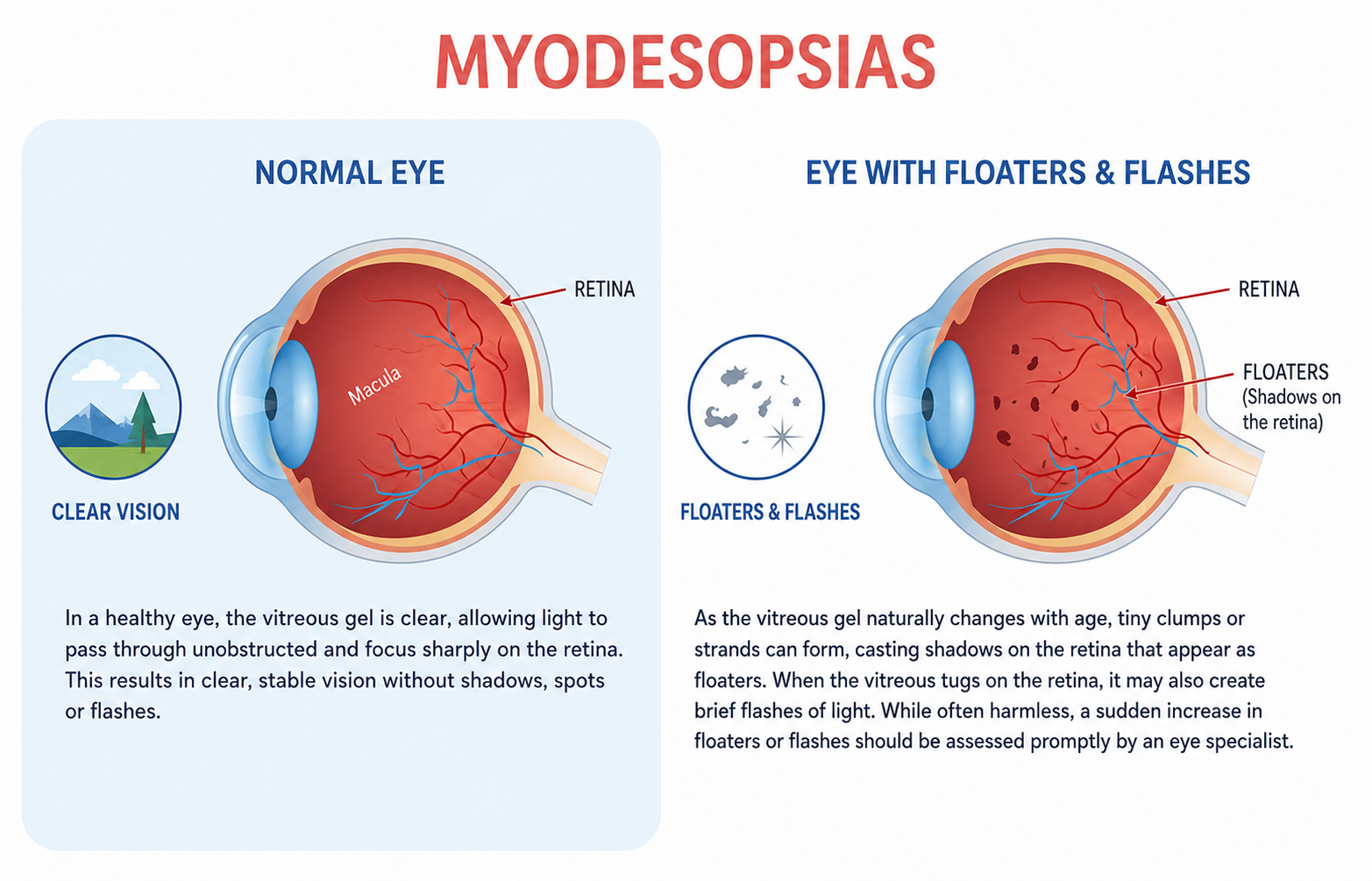
Some patients may experience seeing floating particles in their vision, often described as small moving spots, threads, cobweb-like shapes, rings or shadows. These eye floaters are usually more obvious when observed against a white wall or bright sky. They represent shadows cast onto the retina by the lining of the vitreous gel that has detached from the retina. The vitreous gel is what fills the core of the eye, and the natural ageing process can lead to posterior vitreous detachment. This can also be precipitated by eye trauma.
During the active stage of posterior vitreous detachment, some patients may experience seeing flashing lights at the corner of their eyes. This is due to the pulling forces on the retina and retinal blood vessels from the detaching vitreous. Typically, the flashes will subside after a few weeks, when the posterior vitreous detachment is complete.
Why See a Doctor for Floaters?
Long-standing floaters and flashes that have not changed are generally harmless but they can be bothersome. However, a sudden increase in floaters, or floaters with flashing lights should be assessed by an eye specialist because the same posterior vitreous detachment that produces them can also tug hard enough on the retina to create a retinal break. A break can be sealed with laser retinopexy, but if it progresses to a retinal detachment, vision can be permanently lost without prompt surgical repair.
Floaters can also signal bleeding inside the eye (vitreous haemorrhage) or inflammation. About 2% of patients experiencing posterior vitreous detachment go on to have a retinal break, which is why all patients experiencing new floaters and flashes should have a full eye check with dilated retinal examination to rule out a retinal tear or detachment.
Common Causes of Floaters
Floaters develop when something disturbs the vitreous gel inside the eye. Several conditions can lead to this, from age-related changes to underlying eye disease.
Age-Related Vitreous Changes
The vitreous gel inside the eye gradually liquefies and shrinks with age. As it pulls away from the retina, a process called posterior vitreous detachment (PVD), it can produce floaters.
Inflammation Inside the Eye
Uveitis is inflammation inside the eye that can affect the area behind the lens. It releases inflammatory cells into the vitreous, which appear as floaters. Underlying causes include infection and autoimmune disease.
Bleeding Inside the Eye
Vitreous haemorrhage refers to blood leaking into the vitreous. It can be caused by retinal tears, diabetic retinopathy, or injury. The red blood cells suspended are seen as floaters.
Eye Injury or Previous Eye Surgery
A direct injury to the eye can disturb the vitreous and produce floaters. Intraocular procedures, such as cataract or retinal surgery, may also trigger them, sometimes years after the operation.
When Floaters Need Urgent Attention
Most floaters are not a cause for concern, but sudden changes should be assessed promptly. If any of the symptoms below appear, contact an eye clinic without delay, before vision is permanently affected.
| Sudden increase of new floaters | Persistent flashes of light | Shadow or curtain across vision |
| New floaters after eye surgery or injury | Sudden loss of vision | Blurred vision |
How Floaters Are Diagnosed
A floater assessment begins with your symptom history, followed by a vision check and a dilated retinal examination where the ophthalmologist can inspect the retina and vitreous in detail. If bleeding makes it difficult to examine the retina clearly, an ultrasound scan of the eye may be used. The aim is to rule out a retinal tear or detachment and to judge whether any treatment is actually needed.
Floaters Treatment Options
Treatment depends on what is causing the floaters and how much they interfere with your vision.
When intervention is needed, the options range from laser procedures to surgery, depending on whether the issue is the floaters themselves or an underlying retinal problem.
Monitoring
For floaters that are harmless and stable, a watch-and-review approach is generally appropriate.
Vitrectomy Surgery
A micro-incision day procedure performed under local anaesthesia. The vitreous gel and its debris are removed and replaced with a saline solution. The procedure carries a risk of retinal tears, retinal detachment, and earlier cataract formation.

Laser Retinopexy for Retinal Tears
When a retinal tear is found, a laser is used around its edges to spot-weld the retina, forming a barrier that helps prevent the tear from progressing to a retinal detachment.
YAG Laser Vitreolysis
A focused laser is directed at a large floater, fragmenting it into small pieces. Results vary between patients, multiple sessions may be needed, and there is a risk of retinal injury from the laser energy.
Scleral Buckling for Retinal Detachment
A soft silicone band is fixed to the outer wall of the eye to reposition the retina. The tear is then sealed with freezing treatment. It is performed under local or general anaesthesia.
Floater Removal Surgery and Laser Options
| Option | When it may be considered | Key limitations and risks |
| Laser vitreolysis | May be considered for selected floaters that are suitable in size, position, and distance from sensitive eye structures. | The trade-off is unpredictability. While it aims to fragment a floater, not every floater responds to laser, which means that the result can be hard to forecast, and certain floater types are not suitable for the procedure to begin with. |
| Vitrectomy | May be considered when floaters are persistent, visually significant, and affecting daily activities despite observation. | As an intraocular operation, it sits at the invasive end of the scale. Therefore, recovery may take longer, and the risk profile is correspondingly broader. |
When floaters are interfering with vision, two interventional routes can be considered: laser vitreolysis and vitrectomy. Each option has its own suitability factors, limitations, and risks, so the decision should be based on the patient’s eye condition, floater characteristics, symptom severity, and findings from the eye examination.
For both procedures, the threshold for recommending treatment is set deliberately high. Most surgeons hold off unless the floaters are persistently affecting daily life and vision.
Eye Floater Treatment: What to Expect
Once the assessment is complete, the ophthalmologist explains the findings and walks through the options available. The conversation usually covers the condition of the retina, and which path forward (observation, laser, or surgery) fits the picture. The discussion covers the likely benefits, the limitations, and the risks of each option for your eye.
Risks and Limitations of Floater Removal
No floater treatment can guarantee that every floater will disappear permanently. Some of the limitations include:
- Incomplete improvement: Symptoms may persist after laser vitreolysis, and more than one session may be needed to address the majority of floaters.
- Recurrence: The vitreous keeps changing with age, so new floaters can appear over time even after vitrectomy.
- Procedural risks: Both options carry their own complications, ranging from retinal injury to accelerated cataract formation.
For these reasons, a cautious approach to treating floaters is recommended, reserving intervention for selected cases where vision is seriously affected.
Recovery and Aftercare
Recovery varies depending on the procedure.
After laser vitreolysis, vision usually settles within a day or two, though some patients notice mild floating spots from fragmented particles, which tend to reduce over time.
Vitrectomy involves a longer recovery. The eye can feel sore for the first few days, vision may be blurry while it adjusts, and there are usually restrictions on swimming, strenuous activity, and air travel during the initial healing window. Eye drops are prescribed, and follow-up appointments are scheduled to check the retina and intraocular pressure. The ophthalmologist will advise when normal activity, including driving, can resume.
Our Doctors

Medical Director and Senior Consultant
Ophthalmologist
MBBS (Melbourne), MMed (NUS), FRCS (Edinburgh), FAMS (Ophthalmology)
Subspecialty: Laser Vision Correction and Lens Implant Surgery

Senior Consultant (Visiting)
Ophthalmologist
MBBS (Singapore), MMed(Ophth), FAMS (Ophth),
MCI (Singapore), PHD (Utrecht)
Subspecialty: Retinal Conditions, Floaters & Flashes in Vision, Age-Related Macular Degeneration, Myopia in Children & Adults Cataracts
Payment Options
We accept cash, NETS, and major credit cards.

Interest-Free Instalment Plans
Available with UOB, OCBC, DBS, and POSB bank credit cards.

Why Choose LSC Eye Clinic

RETINAL SUBSPECIALTY ASSESSMENT
Floaters and flashes are reviewed by an ophthalmologist with retinal training, so tears and detachments are not missed.
SATURDAY AVAILABILITY
The clinic opens on Saturday mornings, giving patients an option outside the working week for an eye check.
CENTRAL CLINIC LOCATION
Located at Paragon, Orchard, LSC Eye Clinic is accessible to patients seeking eye assessment and care in central Singapore.
Frequently Asked Questions About Floaters Treatment
Many floaters become less noticeable over weeks to months as they drift out of the line of sight and the brain learns to ignore them. They may not vanish entirely, but they often stop being bothersome without any treatment.
Treat a sudden increase in floaters, repeated flashes of light, or a shadow or curtain across your vision as warning signs. These can point to a retinal tear or detachment and should be examined by an eye specialist.
No. Most floaters are harmless and do not need active treatment. Removal is generally considered only when floaters significantly interfere with vision and after the risks have been discussed with your ophthalmologist.
Laser vitreolysis fragments a floater without incisions. Vitrectomy surgically removes the vitreous gel and its debris.
Yes, floaters can recur. Laser may leave or generate fragments, and the vitreous keeps changing with age. Vitrectomy removes existing debris, though it does not stop all future vitreous-related changes.
Each option carries its own risk profile. Laser and surgery both have potential complications, including retinal injury, which is why treatment is reserved for selected cases and discussed in detail beforehand.
After an eye examination, your vision may be blurry and light-sensitive for a few hours, so arrange alternative transport. After a procedure, your ophthalmologist will advise when it is safe to drive again.
For a floater and retinal assessment, contact lenses do not usually need to be stopped. If other tests are planned, the clinic will let you know in advance whether to attend in glasses.
This depends on the treatment. For instance, monitoring requires no downtime while vitrectomy or retinal surgery needs a longer recovery, which your ophthalmologist will outline.
A sudden increase in new floaters, especially with flashes or a shadow in your vision, should be checked immediately. Contact an eye specialist promptly so the retina can be examined for a tear or detachment.
Other Eye Services

LSC Eye Clinic assesses and treats common eye infections affecting the surface and surrounding tissues of the eye.

The clinic manages pterygium, a growth on the surface of the eye, including assessment and surgical removal where appropriate.