Common Eye Problems and What They Look Like

Although the eye is a small organ, it contains a myriad of even more minute functioning parts that ensure it does its job by giving us one of the most important and beautiful senses of all – sight! When I explain to patients about their eye conditions, I often liken the eye to a camera – not a smartphone camera which is ever so popular these days, but a prototypical 35mm film camera that the baby boomers, Gen X and millennials amongst us would have encountered decades ago!
How do we See?

Vision occurs when light enters the eye through the cornea and the pupil. The cornea is a transparent dome-shaped part of the front of the eye. It is akin to the clear screen in front of the camera lens. The pupil functions just like the aperture in a film camera, controlling the amount of light coming through. Just as how the lens in a camera allows passage of light, the lens in the eye also bends incoming light rays and projects these onto the retina.
The retina is made up by millions of specialized cells known as rods and cones, which work together to transform the image falling onto it into electrical signals. These signals are sent to the optic disc which lies in the central part of the retina, and are then transferred through the optic nerve out of the eye to the brain, where they are processed. It is this combination of millions of individual electrical signals to the brain that constitutes the final perception determining what we see.
Different Parts of Your Eye can be Affected by Different Eye Conditions

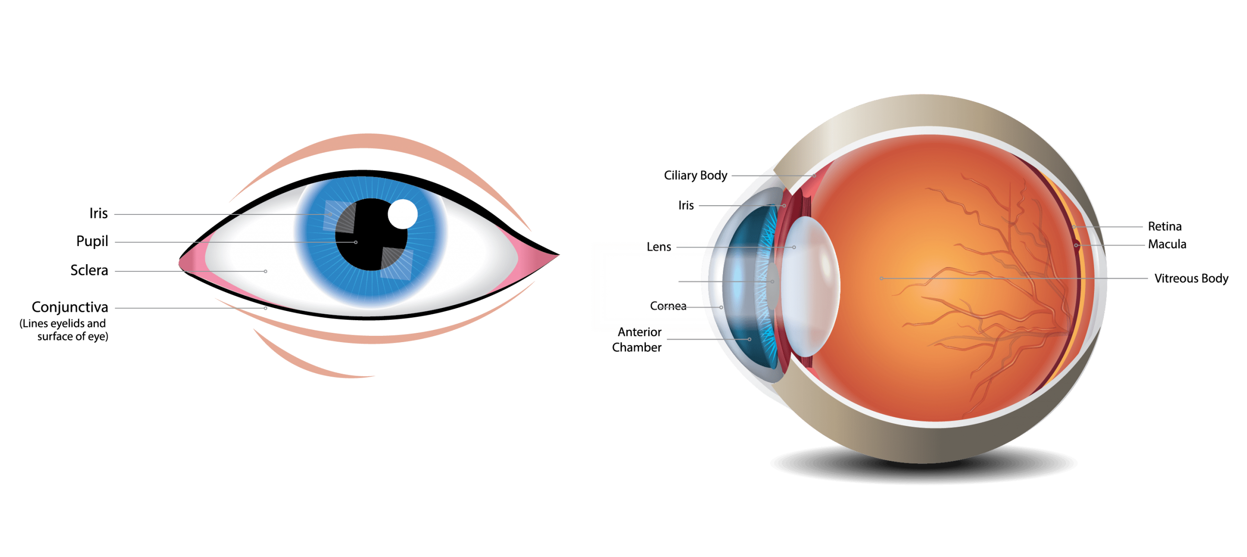
Let’s take a look at the individual structures within the eye, what they do, and what can go wrong with each of these important parts of the eye:
| Parts of the eye | Purpose | What can go wrong? |
|---|---|---|
| Eyelids | The constant blink of the eyelids ensures that the cornea is appropriately coated with a tear film every now and then. | With age, loosening of the supporting tissues of the upper eyelid causes it to droop, leading to eyelid ptosis. When ptosis is severe, vision is affected. However, vision can be restored readily with surgical correction of the ptosis.
Very commonly, a stye or chalazion arises when glands in the eyelids are blocked, leading to a build-up of secretions or oily material. Such conditions can be treated with good outcomes. |
| Conjunctiva | This is a thin, transparent, connective tissue covering enveloping the sclera (i.e., the white of the eye). On its own it is not distinctly visible to an external observer. | It is involved in the very common “red eye”, which is a viral infection of the conjunctiva, as well as in allergic eye diseases. |
| Sclera | The white outer covering of the eyeball, which is continuous with the cornea in the front bit of the eyeball. | Inflammation of the sclera results in a red eye as well, and may appear indistinguishable from conjunctivitis. However, scleral inflammation is much more painful. |
| Cornea | The transparent dome-shaped part of the front of the eyeball. It is an all- important refracting surface, and transmits light entering the eye onto the lens, which in turn focuses it onto the retina | The cornea is sensitive to pain, which is why many corneal conditions like abrasions and ulcers are very painful. Keratoconus is a condition mainly occurring in younger individuals where the cornea becomes progressively weak and irregular, resulting in high levels of myopia and astigmatism.
The cornea is coated and protected by a tear film, and abnormalities in the tear film give rise to dry eye disease. Contact lens overuse also results in tear film abnormalities. Refractive surgeries like LASIK modify the shape of the cornea and alter its refracting properties. |
| Iris | It regulates the amount of light that enters your eye. It forms the coloured, visible part of your eye in front of the lens. Iris color varies depending on ethnicity, and may be green, blue or gray. In Asians, this is usually brown. This shows up visibly as what we commonly refer to as the “dark” part of the eye. | The iris can be affected by inflammation, giving rise to a red and painful eye. Injuries to the eyes may damage the iris as well. In patients with very severe diabetic eye disease, abnormal blood vessels develop on the iris. |
| Pupil | The circular opening in the center of the iris through which light passes into the lens of the eye. The pupil size can change, depending on the amount of environmental light. The pupil enlarges in the dark and narrows in light. In patients with dark irises, it can be difficult to identify the pupil, since the pupil appears black. | Pupils may be abnormally large or small, and if so would have an asymmetric appearance between the 2 eyes.
Such signs may indicate disorders in the nerve-brain pathways and always warrants a need for thorough and semi-urgent evaluation. |
| Lens | A transparent structure situated behind the pupil. It is enclosed in a thin transparent capsule and helps to refract incoming light and focus it onto the retina. | Cataract surgery is the most commonly performed eye procedure worldwide.
A cataract occurs when the lens becomes cloudy, and cataract surgery in Singapore involves the replacement of the cloudy lens with an artificial plastic lens implant. The new lens implant resides within the capsule. |
| Choroid | The middle layer of the eye between the retina and the sclera. It contains a pigment that absorbs excess light to prevent blurring of vision. | The choroid can be involved in inflammatory diseases of the eye. |
| Ciliary body | This structure is continuous with the choroid and the iris. The ciliary body produces aqueous fluid that maintains the volume and pressure of the eye. | Overproduction of fluid by the ciliary body that exceeds the fluid outflow capability of the eye leads to a build-up of eye pressure. Sustained high eye pressures damage the optic nerve and cause glaucoma.
The ciliary body is a muscle from which the lens is suspended. The focussing ability of the lens from distance to near or vice versa is altered when the ciliary body muscle changes its shape. Presbyopia (“lao hua”) occurs when we get older as this muscle weakens and cannot effectively alter the focussing capacity of the lens for near visual tasks, e.g. reading. |
| Vitreous | This maintains a solid gel-like consistency in younger individuals, but softens and becomes more liquefied as we get older. The vitreous gel is naturally stuck onto the retina in the young. As we grow older, bits of solid gel break up, giving rise to the very common visual sensation of “eye floaters”. | The entire degenerative process progresses until the entire vitreous gel body peels away or detaches from the retina. In some of these cases, this may lead to a sudden shower of floaters and flashing lights, because the vitreous detachment leads to bleeding into the vitreous cavity, or results in a retinal tear.
Sudden development of many floaters is an emergency which needs prompt dilated eye examination by an ophthalmologist. If a retinal tear is found, laser treatment is absolutely indicated. |
| Retina | A light-sensitive structure forming the innermost layer of the eye. It is composed of light sensitive cells known as rods and cones. The human eye contains about 125 million rods, which are important for vision in dim light. Cones work best in bright light. There are between 6 and 7 million cones in the eye and they are also essential for distinguishing colors. | Within the retina are numerous blood vessels. In diabetic retinopathy, due to a lack of oxygenation, these blood vessels develop new sprouts of abnormal blood vessels growing into the vitreous, with a high tendency to bleed and scar.
A retinal detachment occurs when the retina peels off the wall of the eye, much like wall paper peeling off the wall. It develops from an untreated retinal tear. This is an emergency that requires prompt treatment to prevent vision loss. |
| Macula | The central-most part of the retina. I consider this the high-definition part of the retina, as this is part is important for transmitting the sharpest and most precise image of light falling onto the retina. | Damage to the macula occurs in age-related macular degeneration when bleeding, leakage and scarring occur.
Epiretinal membranes which form on the macular surface occurs with aging, and causes blurry and distorted central vision. The treatment involves keyhole surgery to remove the membranel. |
| Fovea | The fovea forms a small indentation at the centre of the macula and is the area with the greatest concentration of cone cells. | Macular holes develop at the fovea, affecting the central-most vision. In many cases, retinal surgery is effective in closing holes and improving vision. |
| Optic disc | The optic disc identifies the start of the optic nerve where signals from cone and rod cells leave the eye via the optic nerve to the visual center of the brain. This area is also known as the ‘blind spot’ and everyone has one in each eye! | Glaucoma is the most common cause for damage to nerves at the optic disc.
Early detection and treatment of glaucoma is important to preserve your sight. Optic disc swelling occurs due to a sudden interruption of blood supply to the disc, in inflammatory conditions, or from problems like tumors and bleeds in the brain. |
| Optic nerve | It conveys visual information to the brain. | Patients with diabetes and high blood pressure may suffer an acute interruption of blood supply to the optic nerve, producing a sudden shadow in the visual field.
The optic nerve may be involved in inflammatory disease. |
| Extraocular muscles | These are the “external eye muscles”, which are attached to the external surfaces of the eyeball. There are a total of 6 such muscles, which allow us to move our eyes in many conceivable directions. | Patients with thyroid problems are at a higher risk for thyroid eye disease, which causes inflammation and enlargement of the eye muscles, resulting in a protruding appearance to the eyeballs.
Conditions affecting the brain and nerve connections to these muscles, manifesting as difficulty with eye movements, may signal life-threatening diseases in the brain, and certainly need a thorough evaluation. |
Conclusion
Conditions affecting the eye range from mild disorders creating only a fleeting annoyance, to severe vision-threatening disease, to very rarely life-threatening conditions in the brain.
The following symptoms of eye disease are common and important. All individuals who experience these symptoms should be aware of the need to consult an eye specialist for further evaluation.
- Eye pain
- Red eye
- Teary or itchy eyes
- Blurry vision in central or side vision
- Darkening of vision, even if intermittent
- Floaters
- Flashing lights
- Distorted vision
- Double vision
- Pain with eye movements
- Glare and haloes
- Headaches associated with eye symptoms
- Any abrupt or gradual change in quality of vision, even if not fitting into any of the above symptom descriptions
Some of these symptoms warrant an urgent evaluation by an eye doctor. Other symptoms may be assessed at a non-urgent consultation. It is important to note that early detection and timely and appropriate treatment saves sight.
Call up and speak to us to discuss the urgency of your symptoms, and to schedule an appointment with us.
Request for an appointment today.

Tel: +65 6836 1000
Whatsapp: +65 9843 1000
For general enquiries and appointment only
Email: [email protected]
Monday to Friday: 9am to 6pm
Saturday: 9am to 4pm
*Last Registration Time 2pm
Sunday & PH: Closed


